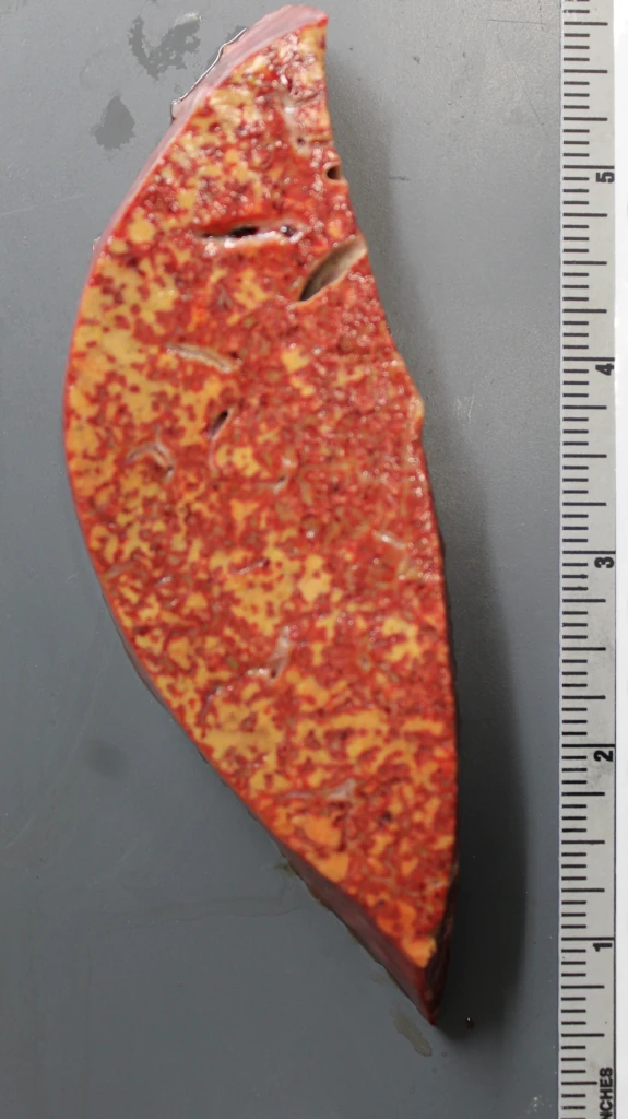
This is a case of a middle-aged woman with diabetes mellitus found unresponsive at home. At the ED, she was noted to have a glucose of greater than 900. She suffered metabolic encephalopathy and multiorgan failure, and died that evening. One prominent finding at autopsy was the presence of innumerable small areas of necrosis in the liver. Here’s a picture of the gross appearance. I apologize for the blurring — the autofocus focused on the ruler and I didn’t notice. Still, it gets the point across:
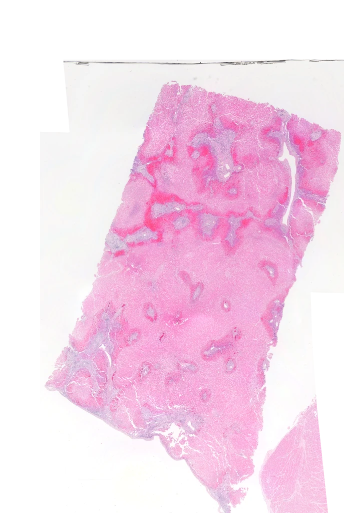
On histology, it turned out that this was probably the worst example of shock liver I’ve ever seen. Normally, of course, shock liver has areas of coagulative centrilobular necrosis with preservation of zone 1 and a good part of zone 2. Not so here. In this case, there’s just a thin rim of viable liver surrounding the portal areas, then the obligatory hyperemia, then coagulative necrosis all the way to the central venule:
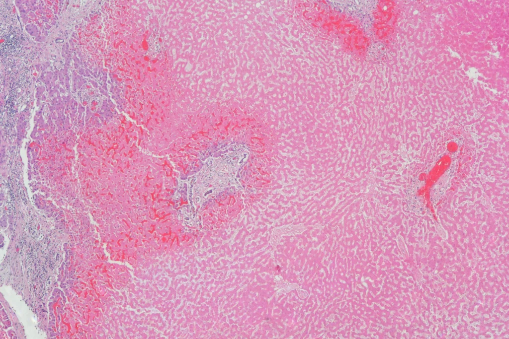
Here’s a medium power view:
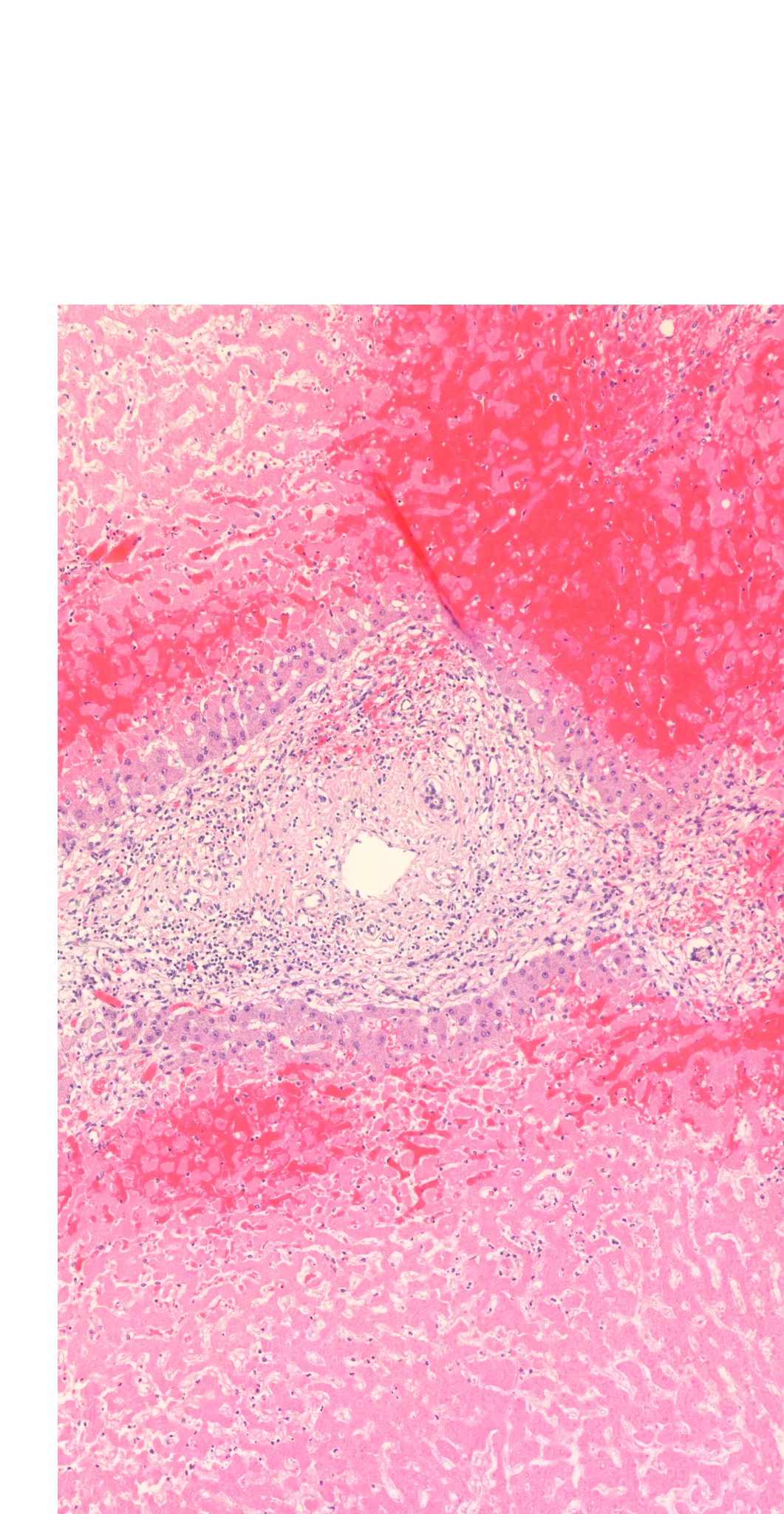
Here’s a higher power, showing the residual viable hepatocytes on right next to the portal area, then hyperemia, then coagulative necrosis with necrobiosis, then just ghost cells as far as the eye can see (off camera to the left):

Here’s a final panorama shot at medium power:
As always, free for use with or without attribution, though attribution is appreciated. If you need high resolution images, let me know.