A 78-year-old man with a complex medical history based primarily in obesity and metabolic syndrome was found dead in bed. He was brought in because of allegations of elder abuse. Autopsy confirmed his multiple comorbidities and no injuries. In addition, his lungs were somewhat firm, resistant to the knife, and very “gritty” when palpated.
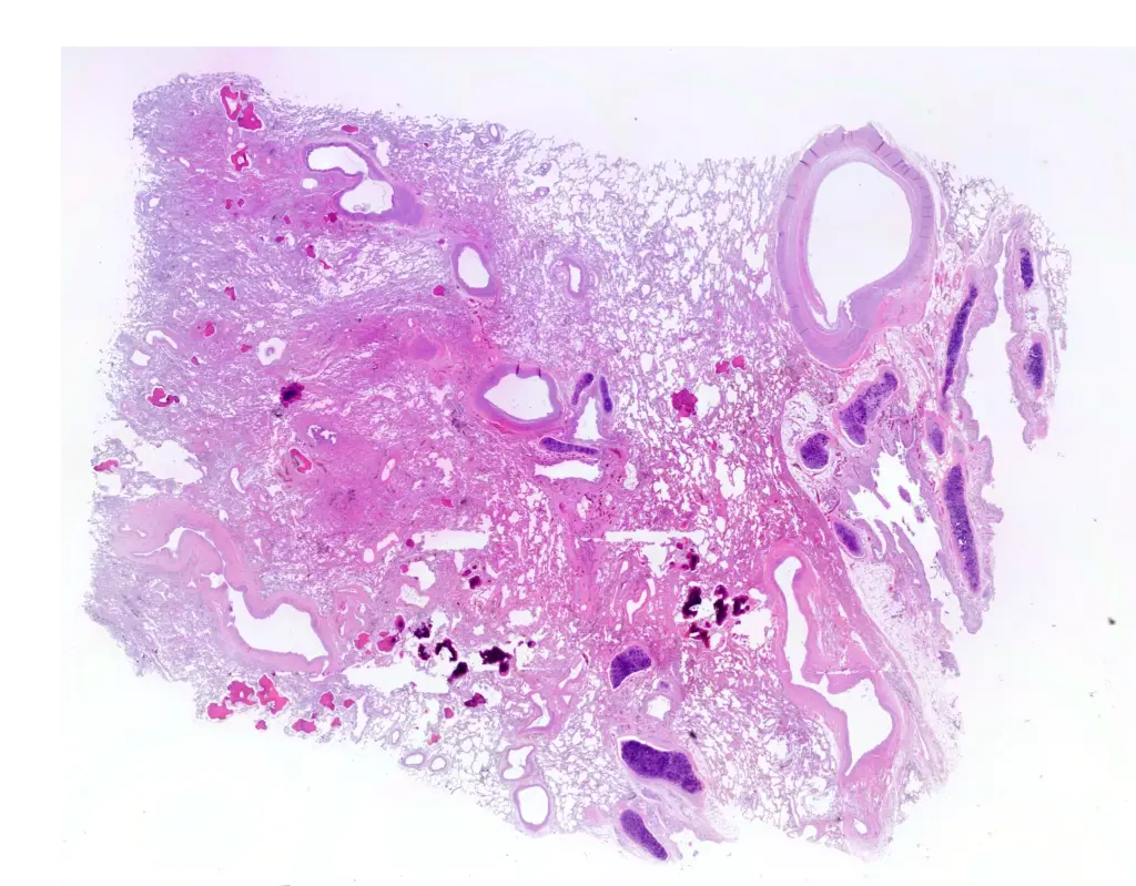
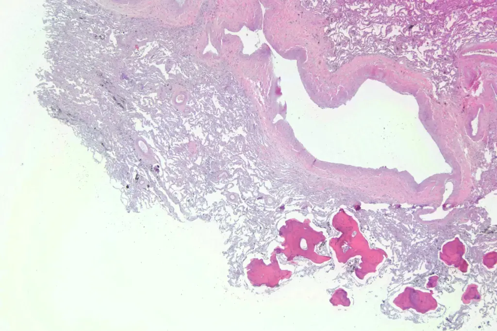
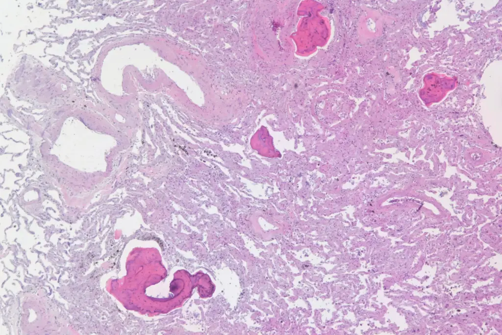
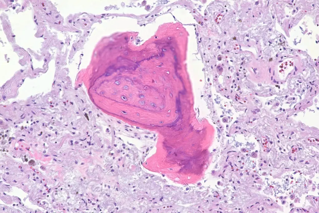
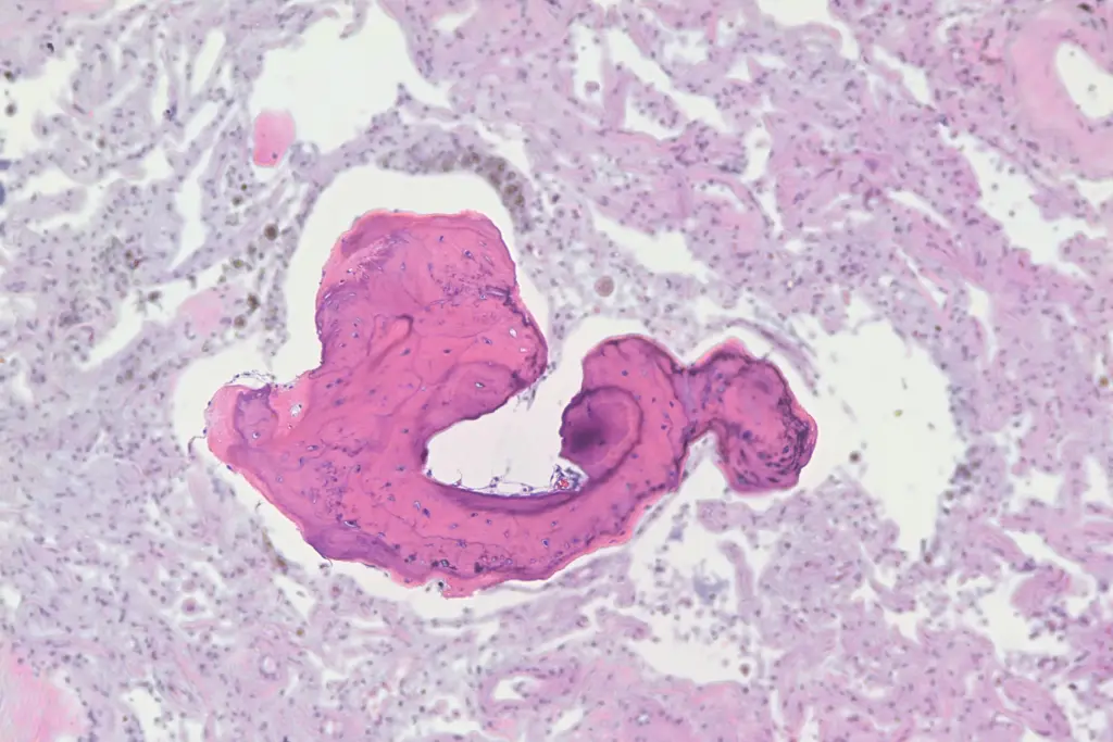
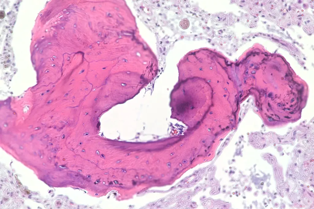
On histology, many of his alveoli were filled with ectopic bone. Ectopic bone formation in the lung is uncommon and is usually asymptomatic, though it can present with symptoms similar to interstitial lung disease. It comes in two types, “dendritic” in which there are branching tree-like shape in small airways, and “nodular” in which small fragments of bone are usually found in alveoli. This is an example of diffuse nodular pulmonary ossification.
Pulmonary ossification can be ideopathic or secondary. Ideopathic cases are more often associated with inflammation. Secondary pulmonary ossification is associated with a myriad of diseases. I don’t understand the mechanics of the pathogenesis, though it’s easy to speculate on minor injury plus calcium metabolism problems. But I don’t know.
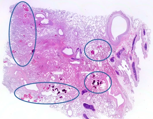
Here’s a panorama of the whole section. You can see the fragments of bone as dark pink objects. In the second image, the more prominent collections are circled:
Here are some higher magnification images:
As always, these images are free to use with or without attribution, though attribution is appreciated. Higher resolution images are available if you contact me.
