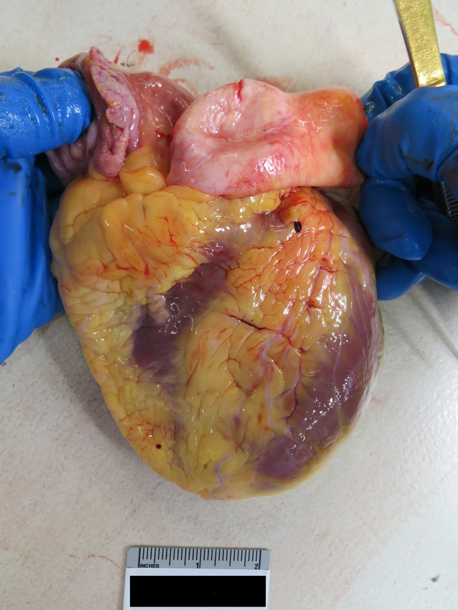
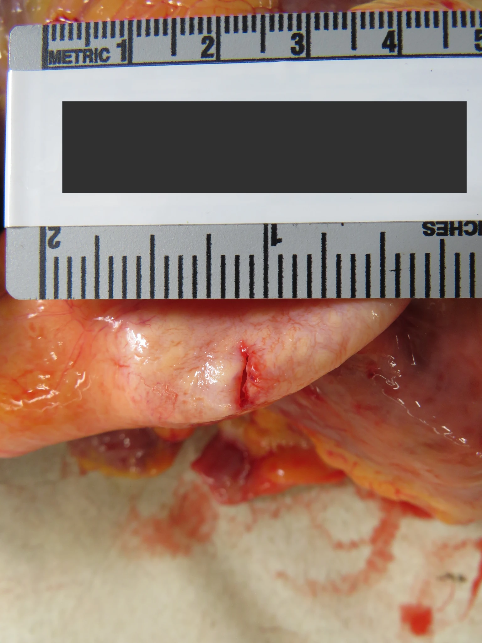
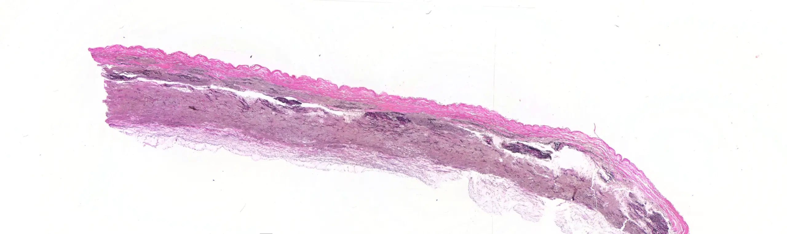
This is a case of a 35-year-old-man found dead at home. His only known medical history was repair of a fractured finger from falling from a bicycle at the age of 32. His blood pressure was elevated at that time, but he was noted to be in pain. At autopsy there were 380 cc of liquid blood in the pericardial space. The heart was not enlarged, but there was an obvious area of balooning and thinning of the proximal ascending aorta, with perforation. The diameter of the aorta at the site of perforation was 4.3 cm, while the diameter immediately above it was 2.1 cm:
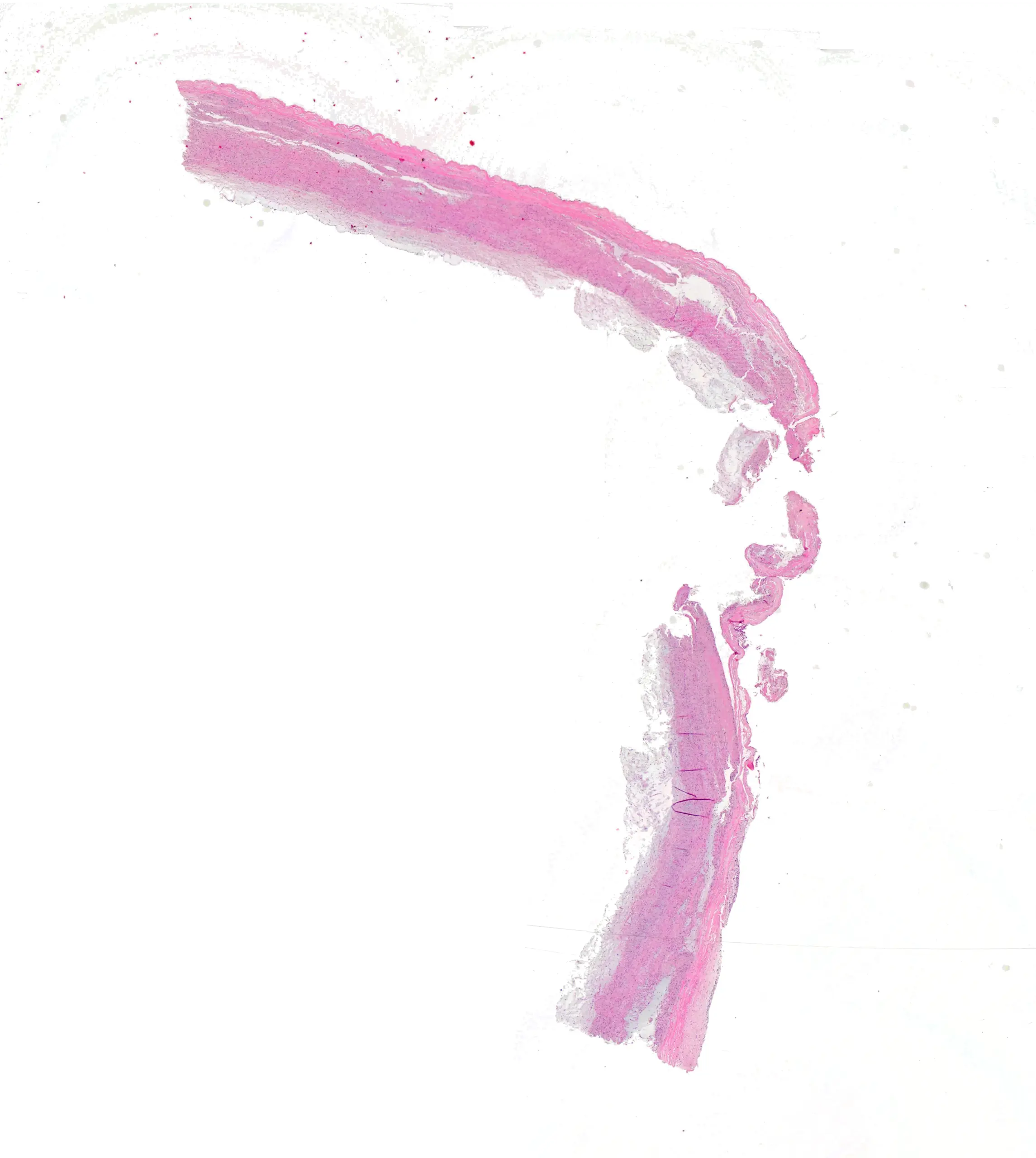
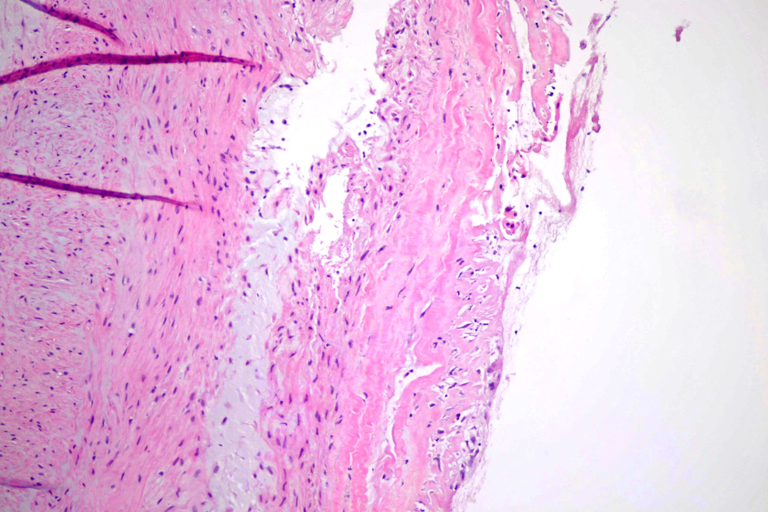
On histologic examintion, it shows the classic findings of these aneurysms. There was notable medial degeneration. Here’s a section at the perforation:
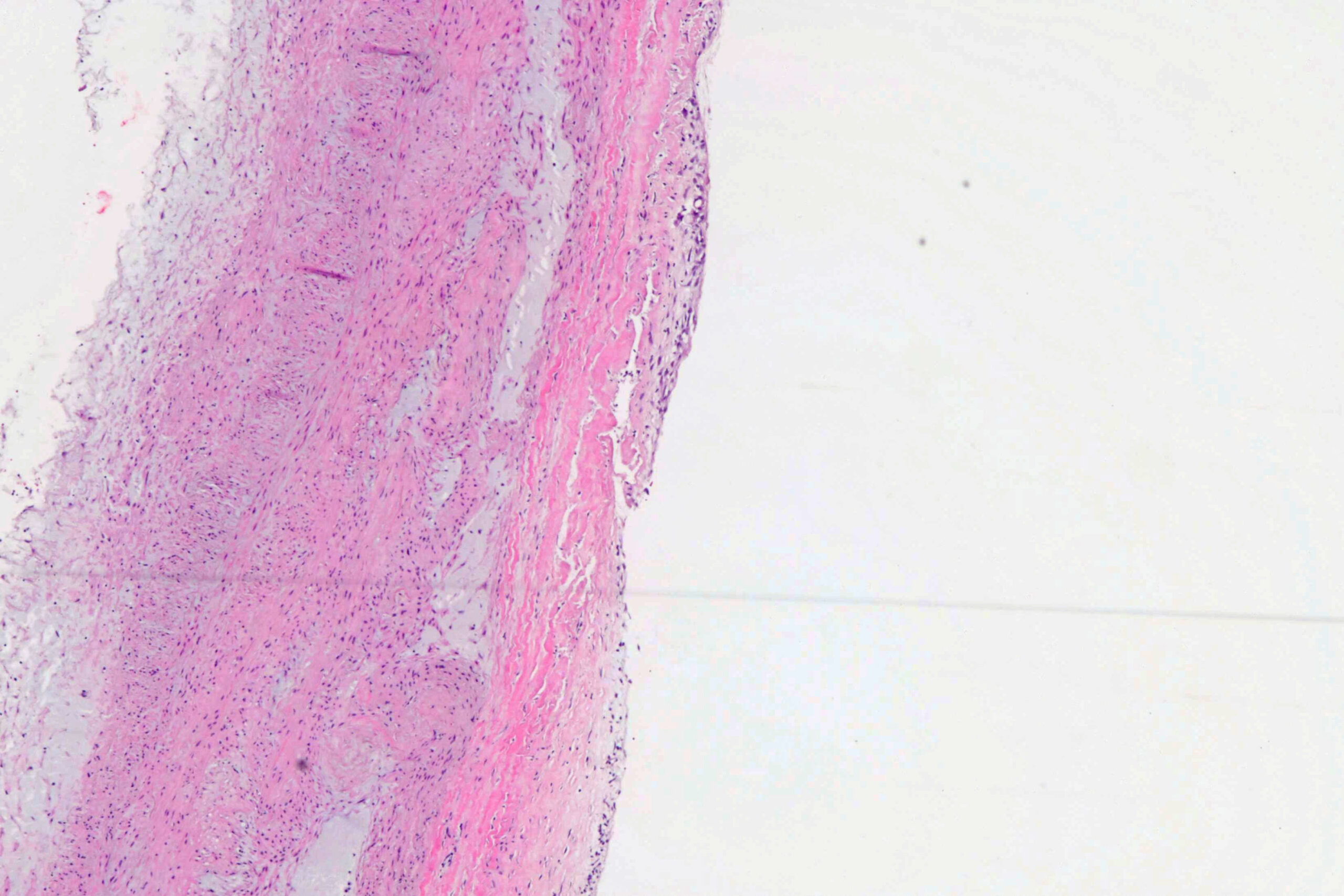
The medial degeneration has the classic blue myxomatous appearance we all know and love:

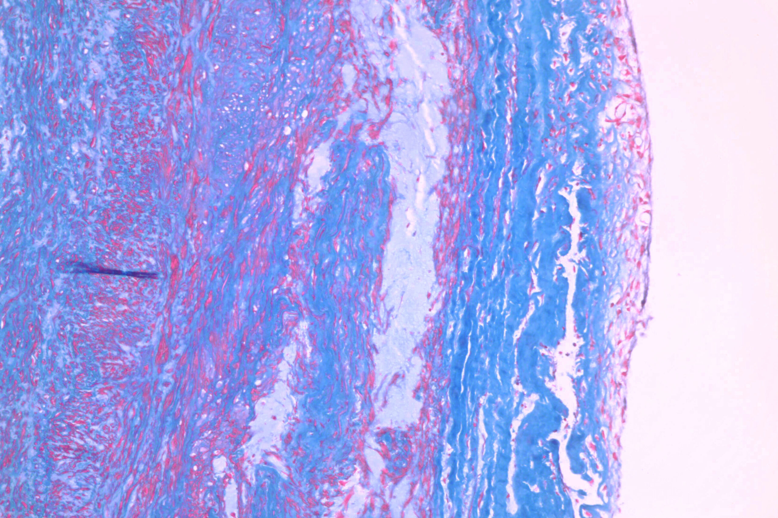
Here’s a trichrome, because it’s pretty:
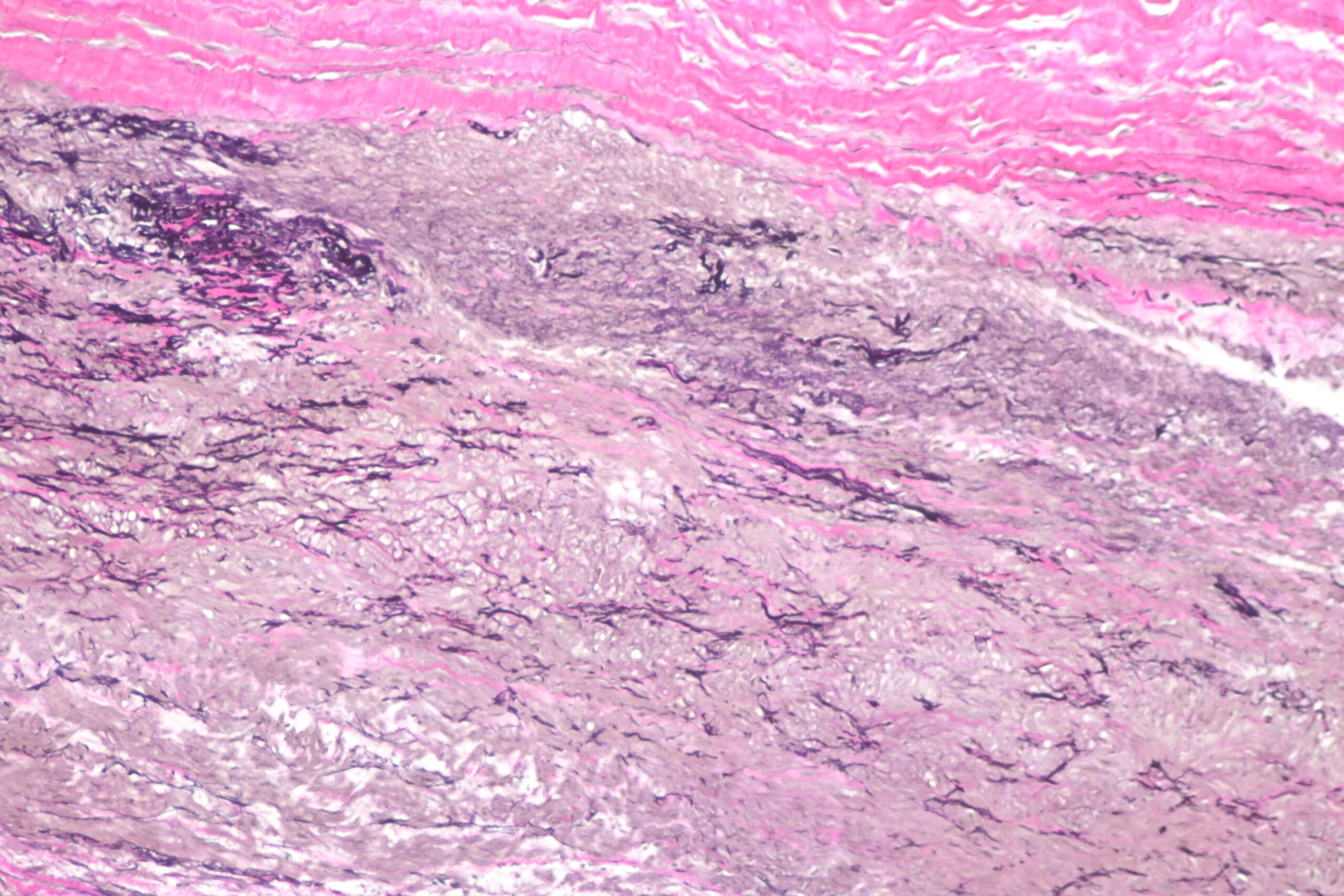
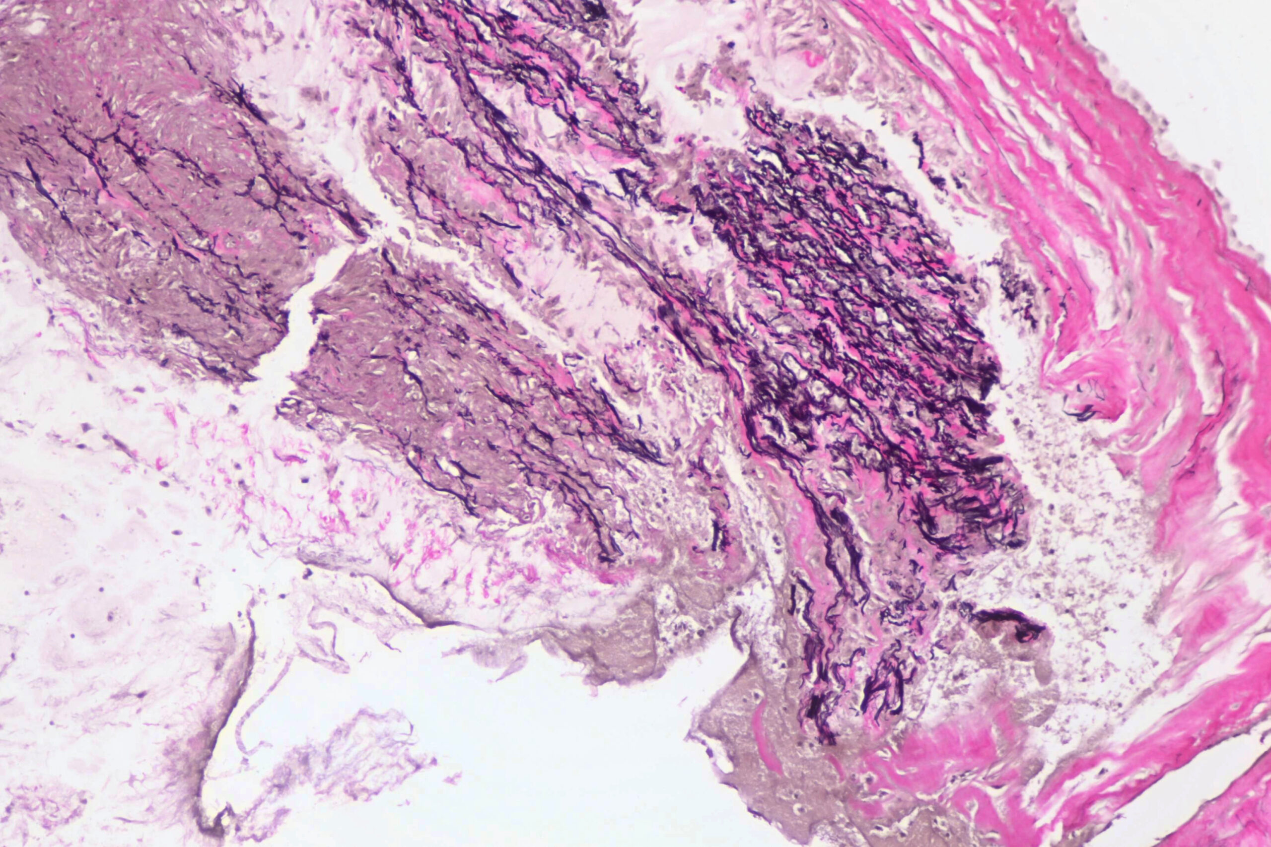
Elastic stain shows classic degeneration and breakup of the elastic lamellae:

There’s a good review of grading of these things at:
Bruijn LE, van Stroe Gómez CG, Curci JA, Golledge J, Hamming JF, Jones GT, Lee R, Matic L, van Rhijn C, Vriens PW, Wågsäter D, Xu B, Yamanouchi D, Lindeman JH. A histopathological classification scheme for abdominal aortic aneurysm disease. JVS Vasc Sci. 2021 Oct 7;2:260-273. doi: 10.1016/j.jvssci.2021.09.001. PMID: 34825232; PMCID: PMC8605212.
It’s open access on PubMed.
As always, free for use with or without attribution, though attribution is appreciated. Higher resolution images available if you contact me.