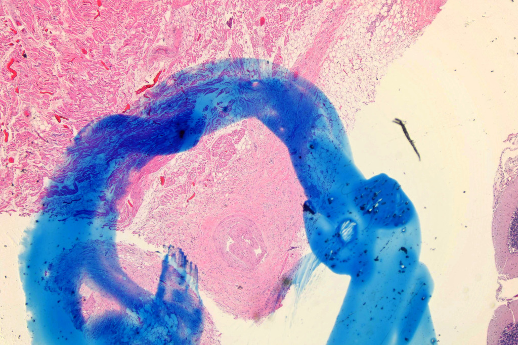
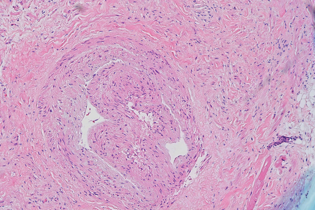
This is the case of a young adult woman with bipolar disorder who came into the ED with nausea, vomiting, and hallucinations. She was given a benzodiazepine and transported to radiology, but arrested on the way. At autopsy, she had no drugs in her antemortem sample, and low doses of benzos and antipsychotics in the postmortem sample. Gross examination was unremarkable. Histologic examination was remarkable for mild steatosis and scattered fibromuscular dysplasia (yes, I’m that old) of multiple small penetrating arteries in her heart. Of particular interest, there was a dissection of the nodal artery of the sinoatrial node:
You can also see a bit of medial degeneration at 12 o’clock in the image.
I routinely take a couple of section of the sinoatrial node and one of the atrioventricular node on my cases. I’m always a little hesitant to diagnose things like “fibrosis” and fatty infiltration in the sinoatrial node, since that seems to be in the eye of the beholder, but this seems real. The sinoatrial node is a low-probability section if you ignore the fibrosis stuff. In some studies, pathologists find lesions about 75% of the time, but my personal opinion is that’s more a function of enthusiasm than real life-threatening lesions. My bar for calling sinoatrial node lesions in the absence of inflammation is higher, but in the past 10 years, I’ve had diagnostic lesions in five or six cases that had nothing else other than a subtle nodal lesion.
I find the atrioventricular node section more productive. I’ve seen lymphocytic infiltration a few times where it was not present in my sections of the ventricles. The penetrating arteries there also seem to be set up for fibromuscular dysplasia if it’s present. Finally, the subendocardium of the interventricular septum near the septal base is a great place to see contraction bands. I see them a lot there in cases where they aren’t there in the other sections. Plus, it captures a bit of the valve leaflets as well, though I don’t obsess about that unless I see something grossly.
In general, I routinely take eight sections of the heart. I take one from each quadrant of the left ventricle (including IVS), one from the right ventricle, one from the atrioventricular node, and two from the sinoatrial node. Why so many? Because as a forensic pathologist, I’m particularly interested in sudden death, and the heart is where the money is on sudden death. Doing each quadrant gives me a survey of vascular distribution. Also, as we all know, acute myocarditis can be very sparse sometimes. I’ve had a lot of cases where I’ve had one small area of lymphocytic aggregation and myocyte necrosis on one slide of the eight. Ischemic lesions are often larger, but are frequently on only one section as well. If I only took one heart section, or just one LV and RV section, I’d have missed a lot of diagnoses over the years.
Update: A colleague brought up a very good point. He said that it looks more like telescoping of the vessel rather than a dissection, particularly since there’s no blood in the media or inflammation. Yeah, that was a concern for me. I brought it to peer review, and the consensus was that this was a dissection. My colleague also brought up the point that blasting the sinoatrial node isn’t usually fatal. You might go into a junctional rhythm or something, but there’s a lot of people walking around with bad upper pacemakers. That’s also true, but there is a literature on sudden death with obstruction of the sinoatrial nodal artery with or without dissection due to fibromuscular dysplasia. These include:
1) James TN, Marshall TK. De subitaneis mortibus. XVII. Multifocal stenoses due to fibromuscular
dysplasia of the sinus node artery. Circulation. 1976 Apr;53(4):736-42. doi: 10.1161/01.cir.53.4.736. PMID:
1253398.
2) Nichols GR 2nd, Davis GJ, Lefkowitz JB. Sudden death due to fibromuscular dysplasia of the sinoatrial
nodal artery. J Ky Med Assoc. 1989 Oct;87(10):504-5. PMID: 2809405.
3) Jing HL, Hu BJ. Sudden death caused by stricture of the sinus node artery. Am J Forensic Med Pathol.
1997 Dec;18(4):360-2. doi: 10.1097/00000433-199712000-00009. PMID: 9430288.
4) Mogler, C., Springer, W. and Gorenflo, M., 2016. Fibromuscular dysplasia of the coronary arteries: a
rare cause of death in infants and young children. Cardiology in the Young, 26(1), pp.202-205
So, I’m sticking with this diagnosis, but I’ll be the first to admit that it’s not with absolute certainty in this case.

