A 35-year-old woman in a home for those with mental disabilities complained of a severe headache. She became unresponsive about 20 minutes later. She was taken to the hospital. CT showed basilar subarachnoid hemorrhage and further evaluation revealed a right PICA berry aneurysm. She underwent emergent embolization, but declined. An autopsy was ordered in the public interest because of an unexpected death in an institutionalized subject.
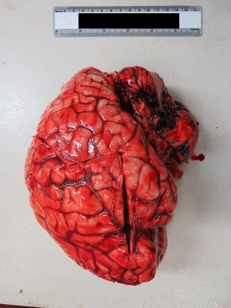
At autopsy, she was noted to have the classic basilar SAH of the perforated berry aneurysm:
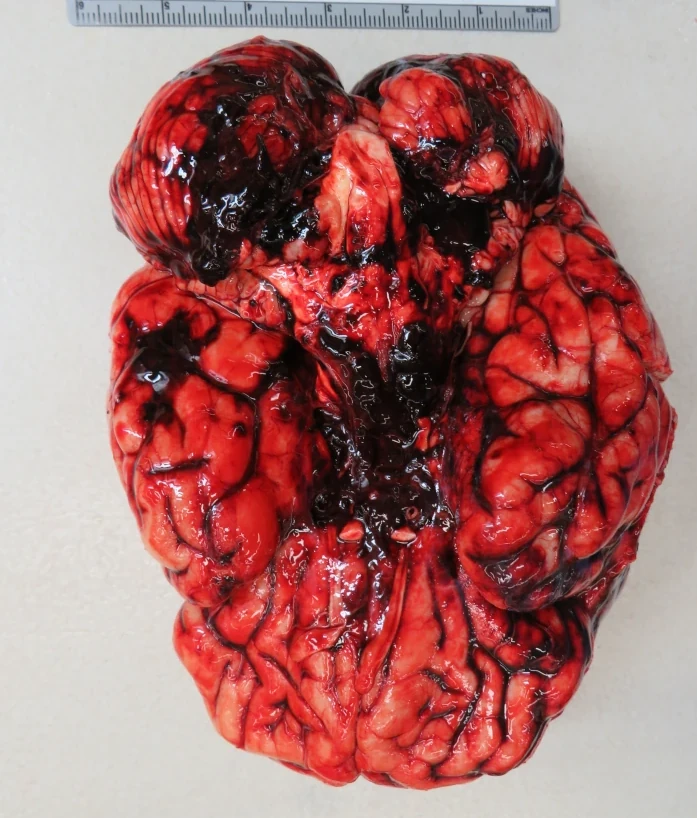
There was also a fair amount of SAH over the left lobe of the cerebellum, and pretty obvious cerebral edema. The left lobe of the cerebellum was soft an necrotic:
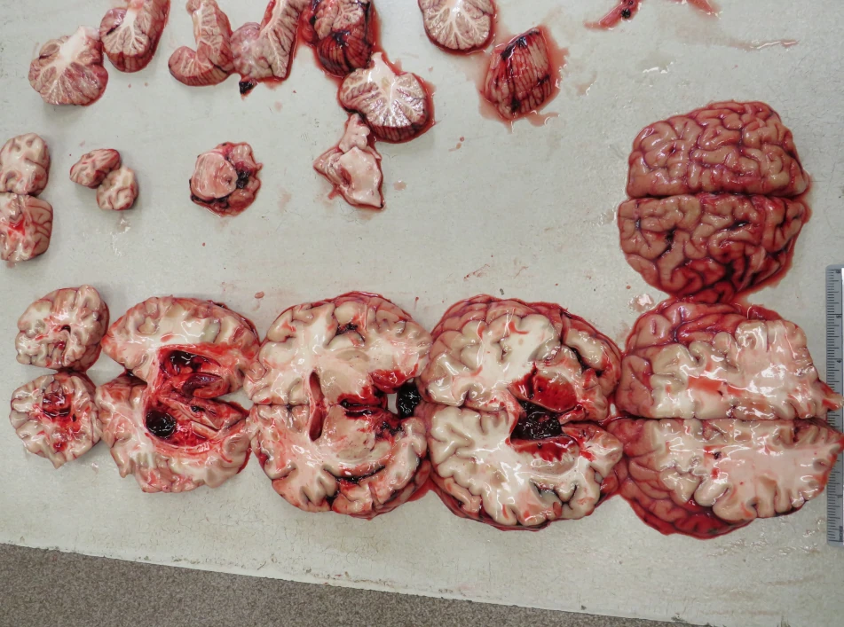
There was hemorrhage into the lateral ventricles, as well as the 3rd and 4th ventricles (yes this is cut fresh — deal with it):
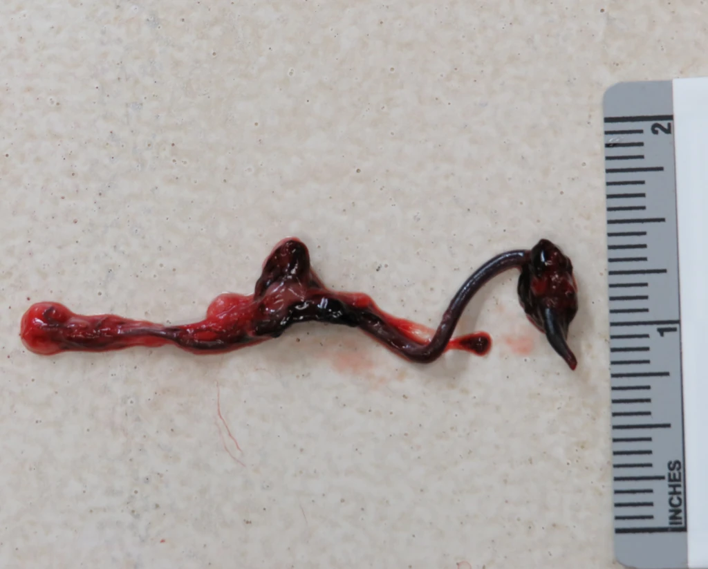
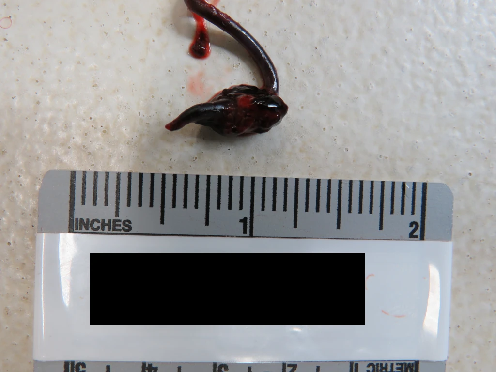
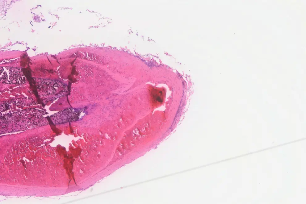
The Circle of Willis looked OK. The left PICA was dark red, extraordinarly firm, and distended. It had a football-shaped aneurysm:
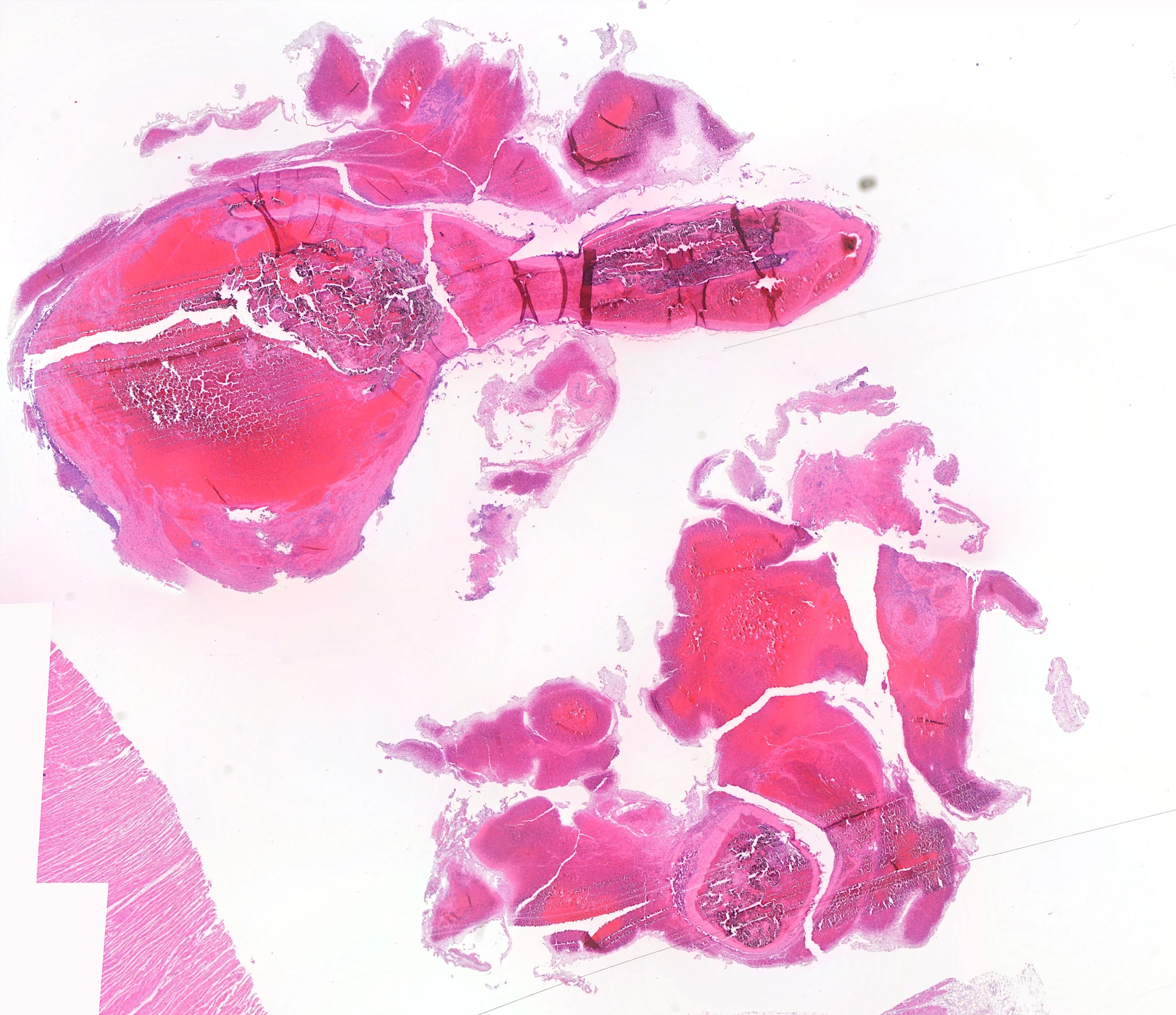
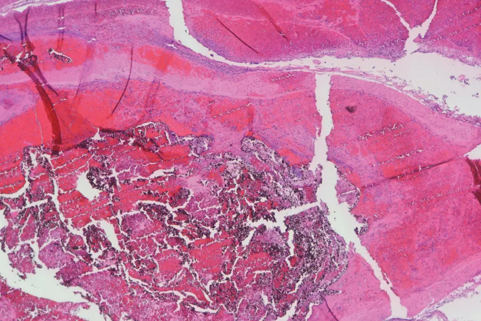
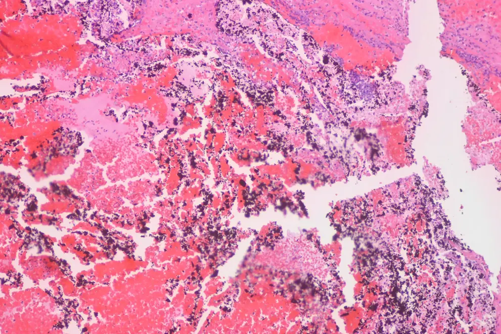
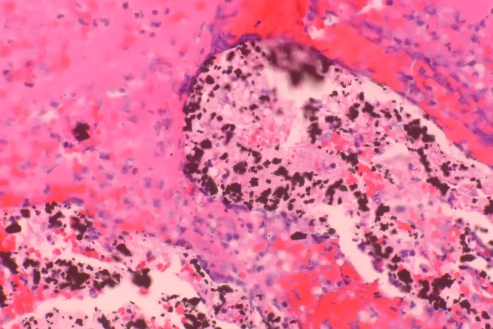
On histologic examination, it was full of black gritty material on H&E. I presume this is the embolization material. There was obvious layered fibrin and red cells surrounding the lumen:
There was a bit of a dissection:
Better seen on elastic stain:
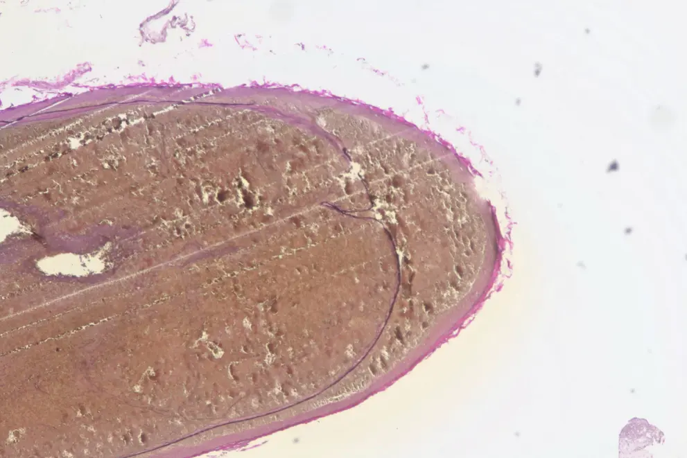
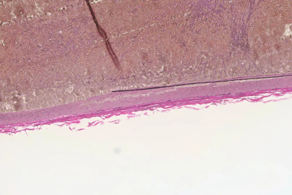
Usually the internal elastic lamina is ratty, but in this case, it just stopped:
Here’s a couple of trichromes because they are pretty (and show the fibrin deposition nicely):
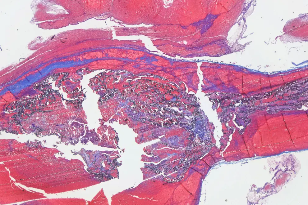
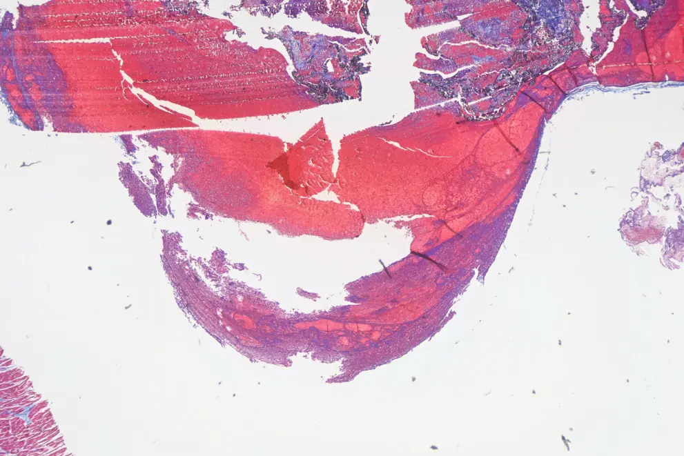
As always, free for use with or without attribution, but attribution is appreciated. If high resolution images are needed, contact me.